Arteries Diagram Labeled | The human heart continues to pumps liters of blood throughout the body all lifelong. Includes an exercise, review worksheet, quiz, and model drawing of an anterior vi The heart is a muscular organ in humans and other animals, which pumps blood through the blood vessels of the circulatory system. You must have seen the human heart diagram. Feb 24, 2021 · function and anatomy of the heart made easy using labeled diagrams of cardiac structures and blood flow through the atria, ventricles, valves, aorta, pulmonary arteries veins, superior inferior vena cava, and chambers.
Blood goes from the left atrium to the left ventricle via the bicuspid (or mitral) valve. Spend a few minutes analysing the diagram, and trying to connect the location of the structures with what you've learned in the video. The femoral artery gives off the deep femoral artery or profunda femoris artery and descends along the anteromedial part of the thigh in the femoral triangle. Thus, you will be aware of the anatomy of the human heart. The strongest muscle in the human body is the human heart.

Two tympani, or … continue reading frog dissection Jul 30, 2021 · a heart diagram labeled will provide plenty of information about the structure of your heart, including the wall of your heart. Feb 24, 2021 · function and anatomy of the heart made easy using labeled diagrams of cardiac structures and blood flow through the atria, ventricles, valves, aorta, pulmonary arteries veins, superior inferior vena cava, and chambers. In human anatomy, the internal thoracic artery (ita), previously commonly known as the internal mammary artery (a name still common among surgeons citation needed), is an artery that supplies the anterior chest wall and the breasts. Jul 27, 2021 · arteries of the cardiovascular system diagram practice test. The femoral artery is a large artery in the thigh and the main arterial supply to the thigh and leg. Blood goes from the left atrium to the left ventricle via the bicuspid (or mitral) valve. Thus, you will be aware of the anatomy of the human heart. You must have seen the human heart diagram. Spend a few minutes analysing the diagram, and trying to connect the location of the structures with what you've learned in the video. In this diagram of the cardiovascular system, you can see labeled structures. If not, you can have a look at the labeled diagram of the human heart present in this article. On the outside of the frog's head are two external nares, or nostrils;
Jul 27, 2021 · arteries of the cardiovascular system diagram practice test. You must have seen the human heart diagram. If not, you can have a look at the labeled diagram of the human heart present in this article. The human heart continues to pumps liters of blood throughout the body all lifelong. The femoral artery is a large artery in the thigh and the main arterial supply to the thigh and leg.
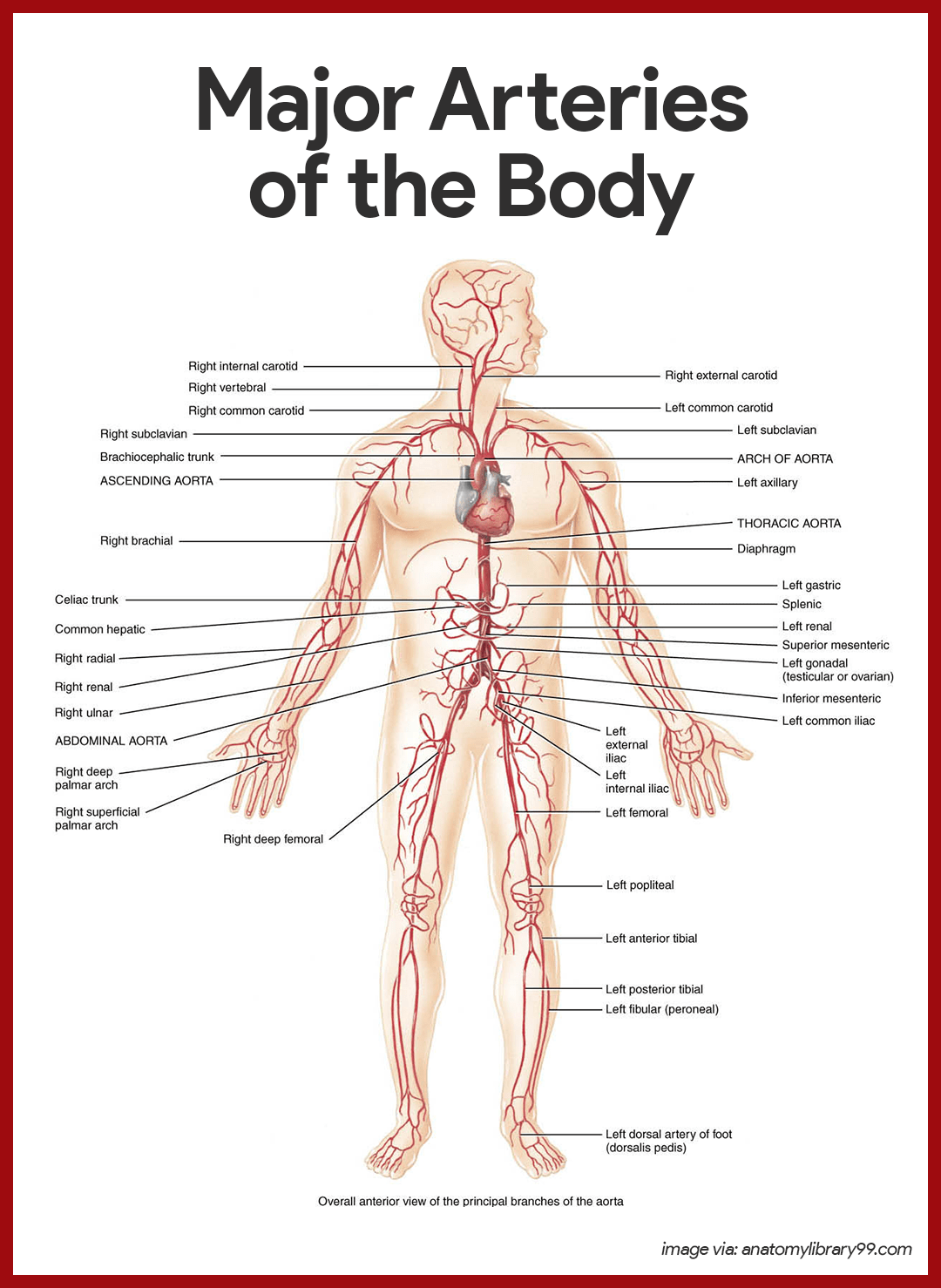
Only one of these is visible on the diagram, a tiny vessel on the right side. The femoral artery is a large artery in the thigh and the main arterial supply to the thigh and leg. Blood then flows through the pulmonary arteries to the lungs where it is oxygenated and then returns from the lungs to enter the left atrium via four pulmonary veins. The wall of the heart has three different layers, such as the myocardium, the epicardium, and the endocardium. In this diagram of the cardiovascular system, you can see labeled structures. Blood provides the body with oxygen and nutrients, as well as assists in the removal of metabolic wastes. Thus, you will be aware of the anatomy of the human heart. On the outside of the frog's head are two external nares, or nostrils; Spend a few minutes analysing the diagram, and trying to connect the location of the structures with what you've learned in the video. Two tympani, or … continue reading frog dissection In human anatomy, the internal thoracic artery (ita), previously commonly known as the internal mammary artery (a name still common among surgeons citation needed), is an artery that supplies the anterior chest wall and the breasts. If not, you can have a look at the labeled diagram of the human heart present in this article. Includes an exercise, review worksheet, quiz, and model drawing of an anterior vi
In this diagram of the cardiovascular system, you can see labeled structures. In human anatomy, the internal thoracic artery (ita), previously commonly known as the internal mammary artery (a name still common among surgeons citation needed), is an artery that supplies the anterior chest wall and the breasts. Here's more about these three layers. Two tympani, or … continue reading frog dissection The strongest muscle in the human body is the human heart.
On the outside of the frog's head are two external nares, or nostrils; The heart is a muscular organ in humans and other animals, which pumps blood through the blood vessels of the circulatory system. Here's more about these three layers. In human anatomy, the internal thoracic artery (ita), previously commonly known as the internal mammary artery (a name still common among surgeons citation needed), is an artery that supplies the anterior chest wall and the breasts. In this diagram of the cardiovascular system, you can see labeled structures. Blood provides the body with oxygen and nutrients, as well as assists in the removal of metabolic wastes. Includes an exercise, review worksheet, quiz, and model drawing of an anterior vi Blood then flows through the pulmonary arteries to the lungs where it is oxygenated and then returns from the lungs to enter the left atrium via four pulmonary veins. You must have seen the human heart diagram. Two tympani, or … continue reading frog dissection Spend a few minutes analysing the diagram, and trying to connect the location of the structures with what you've learned in the video. Thus, you will be aware of the anatomy of the human heart. Feb 24, 2021 · function and anatomy of the heart made easy using labeled diagrams of cardiac structures and blood flow through the atria, ventricles, valves, aorta, pulmonary arteries veins, superior inferior vena cava, and chambers.
The femoral artery gives off the deep femoral artery or profunda femoris artery and descends along the anteromedial part of the thigh in the femoral triangle arteries diagram. Sphenoid bone this is a single bone that is situated at the base of the skull in front of the temporal bones and basilar part of the occipital bone.
Arteries Diagram Labeled: The femoral artery gives off the deep femoral artery or profunda femoris artery and descends along the anteromedial part of the thigh in the femoral triangle.


Post a Comment